


Our team’s main objective is the observation of the brain and more specifically the development of tools for the functional and pathological exploration of the brain at the preclinical and clinical levels. With an expertise in optical and nuclear instrumentation, our developments are more specifically focused on the design of innovative tools dedicated to the physiological and pathological characterization of brain tissue. These tools are targeted on the one hand to cancerology (surgical assistance by endomicroscopy and clinical analysis of tumors) and on the other hand to neuroscience (telemetric implantable probes, behavioral neuroimaging). The team relies on an Experimental Biology cell and on the strong technical potential of the laboratory (mainly engineering pole). It also benefits from the exploitation of the multiphoton imaging platform of small animals “PIMPA” which is at the heart of our developments in optics and allows in particular to feed the data bank on gliomas to determine their grade.
Based on these developments, the main objectives of the team are:
(1) To develop innovative and miniaturized instrumentation dedicated to surgical assistance and analysis of brain mechanisms;
(2) to develop multimodal, multiscale and multispectral imaging approaches for cellular, molecular and tissue characterization in neuroscience and oncology
(3) to adapt preclinical imaging in free-moving animals to provide access to behavioral neuroimaging
(4) To help in the understanding of cellular and tissue mechanisms related to cancer and neuroscience (5) To strengthen our data processing methods and integrate them into our instrumental developments (image processing, database processing by machine learning, pharmacokinetics).
These objectives can be broken down into two research areas:
1) IMOP (intraoperative multimodal optical probe) project:
The objective of the IMOP project is to propose a set of tools to assist in the immediate diagnosis and precise delineation of the edges of infiltrating tumors, in real time during surgery. This project is declined according to four axes:
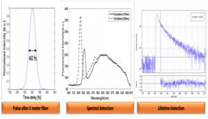
- OGINS: constitutes the main instrumental part of the project and is based on the development of an innovative endomicroscope that can be used intraoperatively and offers a multimodality of contrast response for reproducibility and reliability of the diagnosis in real time.
- Optipen: prototype already developed and based on a bimodal fibered probe, and used in a pilot study at the Paris Psychiatry and Neurosciences University Hospital.
- DATAbank : consists in building a tissue data bank which will be used as a basis for this endomicroscope. It is a multi-scale, multimodal data bank, unique in this field, ranging from deep UV to near IR.
- DATAuse : is directly linked to the database. It classifies the results into groups and subgroups defined with specific thresholds for each type of tissue. This processing of data from the various platforms is based on computer expertise which will probably use the latest advances in AI.
Multimodal detection obtained by the intraoperative endoscope.
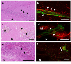
Histological staining (a,c,e) two-photon fluorescence and SHG images (b,d,f) on 3 types of biopsies: control (a,b), metastasis (c,d) and glioblastoma (e,f). scale: a, b 100 µm; c, d, e and f 200 µm
Collaborations: GHU psychiatry and neurosciences and mainly the Neurosurgery and Anatomopathology Department, The Physics of Lasers, Atoms and Molecules laboratory PhLAM – UMR 8523, Lille 1, DISCO line of Synchrotron SOLEIL, The Center of Psychiatry and Neurosciences, IMA BRAIN, INSERM U894, Paris, The BML ” Biophotonics and microsystems laboratory ” of the University of Florida (H. Xi), Beijing institute of technology (School of Information and Electronics), China.
2) MAPSSIC (MAPS-based Intracerebral Probe):
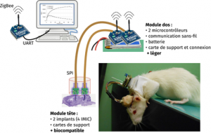
The MAPSSIC project proposes the development of a new generation of radiosensitive intracerebral probes, allowing local concentration measurements of radiolabeled molecules in awake and totally free moving rodents. The goal is to combine molecular data with behavioral data for neuroscience studies, which cannot be done by PET imaging due to the anesthesia or restraint required for the positioning of the animal. We propose to develop a radiosensitive, telemetric and pixelated intracerebral probe based on CMOS technologies to design a low-voltage sensor miniaturized enough to be implantable in a rodent brain. Our ambition is to:
- Design and produce a compact CMOS pixelated sensor compatible with an implantation in a brain tissue
- Design and produce a portable autonomous telemetric electronic device on a rodent
- Evaluate the invasiveness of the probes and its impact on physical and biological measurements over time
- Validate the probes in the context of neurophysiological and neuropathological studies highlighting learning and memory processes, addiction or anxiety in rodents that are awake and free to move.
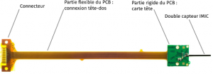
MAPSSIC intracerebral probe made of two IMIC sensors glued back to back, a holding card (rigid part) and a connection card (flexible part).
Collaborations: IPHC Strasbourg, CPPM Marseille, CERMEP Lyon, NeuroPSI Paris-Saclay