


Experimental methodologies:
– In vitro culture: human (BJ, MRI-90, Huvec), rat (RG2, F98) and mouse (3T3)
– 3D cell culture (spheroid, organoid)
– Irradiation of in vitro cultures
– Transfection of plasmid in cell cultures
– Monitoring of cell growth by epifluorescence videomicroscopy
– Epifluorescence and confocal microscopy
– Image analysis and macro realization with ImageJ
– Injection/surgery/ organ removal on rodents
– Transparency of mouse brain
– Sample preparation
– Immunohistochemistry
– Western-blot
– Real time PCR
Equipments :
– 1 cryostat (cold microtome)
– 2 type II microbiological safety stations (PSM II)
– 2 incubators with C02 for controlled cell culture
– 1 inverted microscope for direct observation of cultures coupled to a camera
– 1 epifluorescence video microscope with a thermostatically controlled enclosure with CO2 regulation, for temporal monitoring of growth
– 1 epifluorescence microscope for spatial analysis of biological tissues
– 1 inverted epifluorescence microscope with 37°C chamber for cell culture growth
– 1 microplate reader (fluorescence), for multiple sample analysis
– 1 cell counter (brightfield and fluorescence)

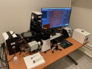
4D videomicroscopy devices for monitoring the growth of living cells for several days (thermostatically controlled chambers).